
If you've ever found yourself pondering what exactly happens during a vein ultrasound, you're in the right place. I'm here to walk you through the intricacies of this fascinating diagnostic procedure, providing insights into why it's done, what it entails, and the valuable information it unveils about your vascular health.
Understanding the Basics: Arteries vs. Veins
There are different types of vascular ultrasound, each serving distinct purposes. One type focuses on examining the arteries in the legs, while the other targets the veins. You might wonder why they need to be done separately, especially if you've recently had an ultrasound of your arteries. The answer lies in the precision of the ultrasound machine, which allows us to examine items that are incredibly close together—mere millimeters apart.
Artery ultrasounds can encompass various areas, such as the carotid artery in the neck and the aorta to check for aneurysms. On the other hand, vein ultrasounds delve into a different realm, seeking to understand whether the veins are functioning optimally. The primary focus is on detecting vein reflux, a condition where blood flows backward, as well as identifying any evidence of old blood clots—both current and past.
The Standing Reflux Ultrasound Exam: Unveiling Abnormal Circulation
Let's start with the reflux ultrasound, a critical component of vein examination. It's important to note that a venous ultrasound must be done standing up rather than supine, or lying down. Standing venous reflux ultrasound is the standard of care because ultrasounds performed lying down very frequently miss the presence of reflux.
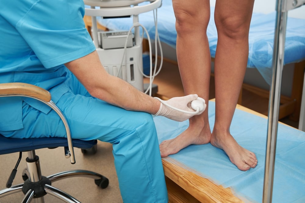
The process is quite simple. You wear a pair of shorts so that your legs are easily seen and available to the ultrasound technician (sonographer). You'll stand on a small stepstool while the technician rolls an ultrasound wand up and down your legs to examine your veins. Tiny ultrasound waves bounce off the tissues beneath your skin, creating a detailed image of the circulatory landscape on the ultrasound screen. The color of the image changes depending on what is happening with the veins - whether blood is pooling or circulating normally.
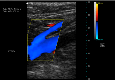
A sample image below shows you an example of what the technician may see on the screen as they hold the ultrasound wand over your lower legs. Blood flow is color-coded and lets the sonographer know what direction the blood is flowing.
Why is it important that this examination is done standing up?
It's because abnormal circulation in veins is most evident when individuals are upright. Gravity comes into play, causing blood to accumulate and pool in the lower legs. For those with a vein condition, a reflux exam while lying down might not reveal abnormalities, but as soon as you stand up, the story changes.
This standing exam is the standard of care in determining the presence of vein reflux, offering a real-time snapshot of how blood circulates when you're on your feet. The precision and accuracy of this procedure are truly remarkable.
The Deep Dive: Identifying Blood Clots in Deep Veins
Now, let's shift our focus to the examination of deep veins for blood clots—a separate aspect of the ultrasound typically conducted while lying down. The sonographer begins the examination just above the groin, meticulously scanning all the way down to the ankle. Specific veins are scrutinized, with a keen eye on detecting signs of acute clots (recent formations) or those that have been present for several weeks or longer.
During this part of the exam, ultrasound clues help unveil the age of the clot, while also highlighting areas where blood flow may be blocked. Lack of blood flow often signals the presence of a blood clot, giving us crucial information about the state of your veins.
This comprehensive examination takes approximately 45 minutes, and the best part? It's entirely painless. The information gathered during this time provides us with a wealth of insights into your vein circulation to establish whether you have a vein condition.
Decoding the Information: From Diagnosis to Treatment
The significance of a vein ultrasound extends beyond the diagnostic realm. It plays a pivotal role in deducing whether the symptoms you're experiencing are linked to a vein condition. If a vein-related issue is identified, the information gathered guides the treatment plan tailored specifically for you.
Every patient is unique, and each vein condition requires a personalized approach. The treatment plan may differ based on the severity of the condition, which veins are experiencing reflux, and the individual goals of the patient.
In summary, a vein ultrasound is not just a diagnostic tool; it is a guide to help us create a plan to treat underlying vein abnormalities. The journey from examination to diagnosis to treatment can only be accomplished with an adequate vein reflux ultrasound.